Research groups
Mouse/rat lines to study sex differences
The following lines are currently available at the University of Oxford:
- Four Core Genotypes (FCG) mouse model
- Sex Chromosome Trisomy (SCT) mouse model
- FCG-like rat model
If you are interested in using these models in your research please get in touch with me.
Kamila Szulc-Lerch
Senior Research Associate
- OxCIN MRI Graduate Programme Deputy Director
- Principal Investigator
I obtained my MSc in Physiology and Neuroscience and PhD in Biomedical Imaging from New York University. My doctoral work focused on developing and applying novel preclinical MRI approaches to studies of brain development using mouse models of human neurodevelopmental disorders. In my studies I used the cerebellum as a model system for quantitative analysis of patterning processes taking place at early postnatal stages.
Following my PhD, I undertook postdoctoral training, funded by a fellowship from Brain Canada and Kids Brain Health Network, at the Hospital for Sick Children and the Mouse Imaging Centre in Toronto. During this period, I worked on projects exploring the potential of pharmacological and lifestyle interventions to promote brain repair in children with brain tumours. I also worked with preclinical rodent models of radiation and hypoxia-ischaemia induced brain injury.
I joined the University of Oxford in the summer of 2019 to build my research programme in preclinical imaging of post-stroke brain recovery. I am principal investigator (PI) on a John Fell Funded study investigating sex differences in stroke recovery using novel Four Core Genotype (FCG)-like rat model. This project is run in collaboration with Art Arnold, Yvonne Couch, Thomas Okell, David Bannerman and Stuart Peirson.
In addition to my preclinical work, I have a strong interest in using big data in clinical neuroscience. I am PI on a UK BioBank project investigating the impact of metformin treatment on brain structure and function. More information about the project can be found here. In collaboration with Novo Nordisk, we have recently also begun examining the impact of obesity, a major risk factor for stroke, on the brain.
POSTDOCTORAL FELLOWS
Quin Yuhui Xie (Novo Nordisk-Oxford Postdoctoral Research Fellow, Oxford Co-supervisors: Jason Lerch and Rogier Mars, Novo Nordisk Co-supervisors: Giorgio Caratti and Chenxi Qin)
D.PHIL STUDENTS
Myrto Lavda (DPhil in Clinical Neurosciences, University of Oxford, Co-supervisors: Yvonne Couch and Heidi Johansen-Berg)
Tien Ho (DPhil in Clinical Neurosciences, University of Oxford, Co-supervisors: Rogier Mars and Daniel Anthony)
M.SC./FHS STUDENTS
Tran Quan (MSc in Clinical and Therapeutic Neuroscience, University of Oxford)
D.PHIL PROJECTS / RESEARCH PLACEMENTS
To inquire about available placements, please email: kamila.szulc-lerch@ndcn.ox.ac.uk and include your CV along with a letter of intent describing your research interests. More information about the DPhil programme in Clinical Neurosciences at NDCN can be found here.
ALUMNI
Nourhane Boudechicha (Visiting Summer Student, MSc in Neuroscience, Aix-Marseille University)
Hannah Davis (FHS, University of Oxford)
Eugenio Graceffo (MSc in Neuroscience, University of Oxford)
Tien Ho (MSc in Clinical and Therapeutic Neuroscience, University of Oxford; *currently DPhil student in Clinical Neurosciences at Oxford)
Emily Moore (MSc in Neuroscience, University of Oxford)
Claire Park (MSc in Neuroscience, University of Oxford)
Tran Quan (Visiting Summer Student, Constructor University; *currently MSc student in Clinical and Therapeutic Neuroscience at Oxford)
Katie Thompson (MSc in Neuroscience, University of Oxford)
David Zavaleta Rivera (Visiting Summer Student, Undergraduate, UC San Diego)
NEWS AND UPDATES
2025 OxCIN/BRC Seed Grants announced
Two of our project were awarded funding to support scanning and research costs:
- Structural correlates of stress resilience in mice
- Effect of rodent brain perfusion fixation protocols on MRI microstructure and histology
2025 Pre-clinical Patient and Public Involvement project
Excited to be part of the wider project team for "Developing resources for public involvement in animal research”.
2024 Vice-Chancellor's Awards
Honoured to have been on the “Your Amazing Brain: A University and Regional Museum Partnership” project team - shortlisted for the Vice-Chancellor's Award in Research Engagement and Highly Commended.
2023 NDCN DEPARTMENT AWARD FOR TEACHING
Grateful to be recognised for contributions to the MRI Graduate Programme with the Departmental Award for Teaching.
Key publications
-
Exercise promotes growth and rescues volume deficits in the hippocampus after cranial radiation in young mice.
Journal article
Szulc-Lerch K. et al, (2023), NMR Biomed
-
Metformin Effects on Brain Development Following Cranial Irradiation in a Mouse Model.
Journal article
Yuen N. et al, (2021), Neuro Oncol
-
Repairing the brain with physical exercise: Cortical thickness and brain volume increases in long-term pediatric brain tumor survivors in response to a structured exercise intervention.
Journal article
Szulc-Lerch KU. et al, (2018), Neuroimage Clin, 18, 972 - 985
-
Mouse MRI shows brain areas relatively larger in males emerge before those larger in females.
Journal article
Qiu LR. et al, (2018), Nat Commun, 9
-
4D MEMRI atlas of neonatal FVB/N mouse brain development.
Journal article
Szulc KU. et al, (2015), Neuroimage, 118, 49 - 62
-
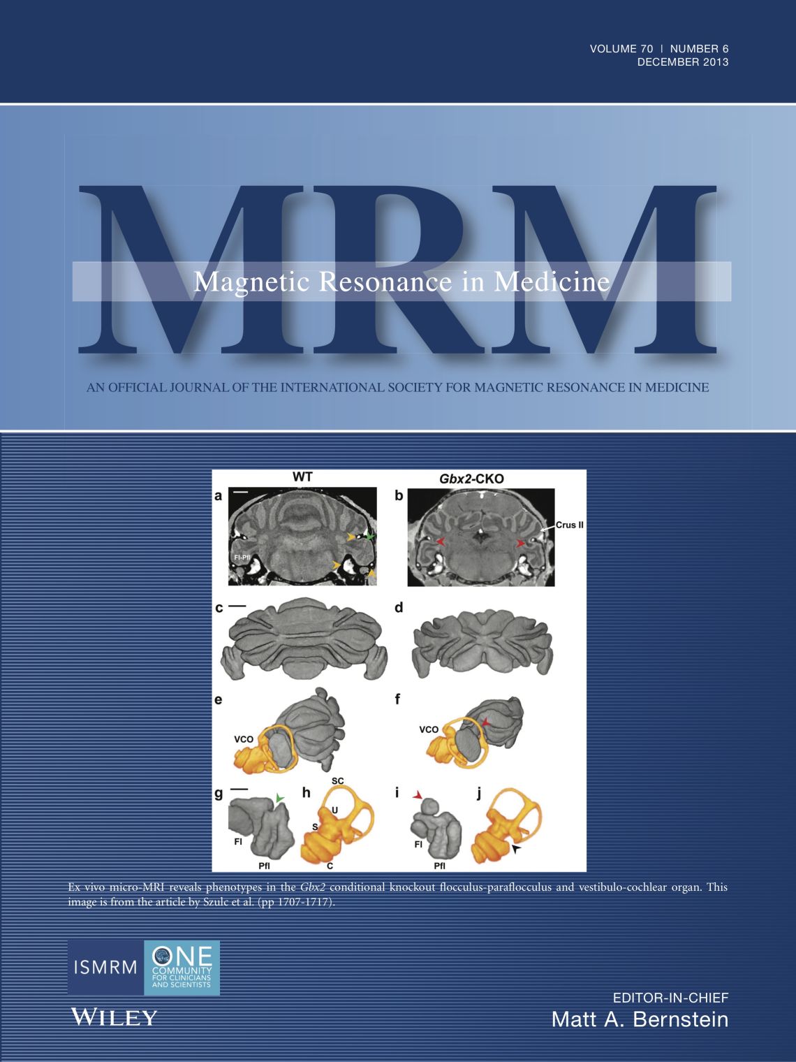
MRI analysis of cerebellar and vestibular developmental phenotypes in Gbx2 conditional knockout mice.
Journal article
Szulc KU. et al, (2013), Magn Reson Med, 70, 1707 - 1717
Recent publications
-
Exercise promotes growth and rescues volume deficits in the hippocampus after cranial radiation in young mice.
Journal article
Szulc-Lerch K. et al, (2023), NMR Biomed
EVENTS OF INTEREST
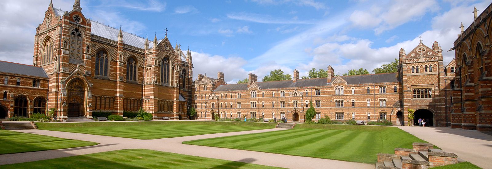
7th UK Preclinical Stroke Symposium, 8 & 9 September 2025, Keble College, University of Oxford (Organising Committee: Yvonne Couch, Paul Holloway and Kamila Szulc-Lerch)

Team Members and Collaborators
-
Myrto Lavda
DPhil Student in Clinical Neurosciences
-
Tien Ho
DPhil Student, Jardine Scholar
-
David Bannerman
Professor of Behavioural Neuroscience
-
Yvonne Couch
Associate Professor of Neuroimmunology
-
Heidi Johansen-Berg
Pro-Vice Chancellor (Strategic Initiatives)
-
Jason Lerch
Professor of Neuroscience
-
Rogier Mars
Professor of Neurosciences
-
Thomas Okell
Associate Professor
-
Stuart Peirson
Professor of Circadian Neuroscience


