©
John Cairns
©
John Cairns
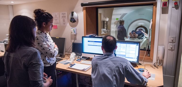
Functional Magnetic Resonance Imaging (fMRI) uses blood flow and oxygen metabolism to infer brain activity. Researchers at FMRIB (now part of the University of Oxford's Wellcome Centre for Integrative Neuroimaging, or WIN) use cutting-edge MRI scanning technology and powerful computational methods to study the human brain in health and disease.
On 20 April 2023 alumni, faculty, and students came together to celebrate FMRIB's 25 years, at an event at the Blavatnik School of Government, University of Oxford. The programme featured reflections on FMRIB's early days as well as its future. It included research highlights on mapping brain function, data analytics, neurophysiology and MR methods. FMRIB alumni spoke about 'Life after FMRIB', and the day concluded with a celebratory drinks reception and buffet.
How did it all begin?
Three University of Oxford professors secured the initial Medical Research Council (MRC) core grant which led to FMRIB's establishment on the John Radcliffe Hospital site back in 1998. Alan Cowey worked in the Department of Experimental Psychology, researching visual neuroscience; John Newsom-Davis was a neurologist who saw the huge potential of MRI for clinical work; and George Radda was a chemist turned physiologist using Nuclear Magnetic Resonance.

Paul Matthews became FMRIB's first Director. He was a neurologist with an interest in Multiple Sclerosis. He was responsible for recruiting the initial FMRIB team including Irene Tracey, Steve Smith, Peter Jezzard and Stuart Clare. FMRIB's focus on balancing methods development with basic and clinical neuroscience applications was core to the original vision.
Expansion over the years
In 2003 FMRIB was physically extended, which allowed the construction of laboratories for additional neuroimaging and stimulation methods including EEG (electroencephalogram), TMS (transcranial magnetic stimulation), and tDCS (transcranial direct current stimulation).
Professor Irene Tracey, now Vice-Chancellor of the University of Oxford, became the second Director of FMRIB in 2005. She studied biochemistry at Merton College before carrying out some of the early studies using fMRI at Harvard University. During her ten-year tenure as its Director, FMRIB was awarded £8 million by the MRC, EPSRC, Wolfson Foundation and University of Oxford to purchase and install new 7T and 3T leading-edge MRI systems. This enabled researchers to image brain structure and function at even higher resolution.

In 2015 Professor Heidi Johansen-Berg was appointed Director of FMRIB and led the bid to bring together FMRIB, the Oxford Centre for Human Brain Activity in the Department of Psychiatry, and the imaging facilities within the Department of Experimental Psychology. The new Wellcome Centre for Integrative Neuroimaging (WIN) launched in 2017.
The Centre Award from the Wellcome Trust, together with generous support from the University of Oxford, allowed the Centre to upgrade and expand its pre-clinical facilities, expand and develop Centre support staff and expand into new office and meeting space.
FMRIB's annexe was completed in 2019. It houses a number of research groups, such as those developing new MRI technologies and analytic approaches, as well as groups investigating how the brain recovers after damage, how the brain processes pain and visual inputs. It is also a space for researchers from across the whole Centre to meet, share ideas and train new researchers.
This expansion also meant that the reception and patient consultation spaces in FMRIB's original facility could be improved. This has resulted in a better experience for the hundreds of research participants who are scanned there every year.
The research and how it's making a difference
WIN brings together neuroscientists, clinicians, and methodologists with a background in physics or image analysis. They use their combined expertise to understand how the brain works, and investigate the underlying causes of conditions such as dementia, psychiatric disorders and vascular disease.
The research at FMRIB/WIN bridges the gap between laboratory neuroscience and human health, by exploiting the capacity of neuroimaging to provide measurements that are sensitive to cellular phenomena and that can also be acquired in living humans. This is achieved by focusing on five themes: Cross-Species Neuroimaging, Cross-Scale Relationships, Population Data Mining, Clinical Markers and Open Neuroimaging.
One of FMRIB's major achievements has been to develop one of the world's most popular software tools for analysing brain images, FSL, which is used in over 5,000 laboratories across the world.
Researchers at WIN are using big data to understand the brain and tackle brain disease. For example, the UK Biobank project is scanning the brains of 100,000 individuals – making it by far the largest brain imaging study in the world. FMRIB researchers have built computer algorithms to mine that data to extract useful measures of brain health and made these available to the research community worldwide. These tools can then be used alongside health data to address questions such as whether brain scans can predict who is likely to get dementia later in life or how COVID-19 changes the brain.

From the very beginning, FMRIB researchers have focused on sharing their work and findings beyond academia, believing that engagement enriches the research, inspires others and builds trust. For example, in 2013, comedian Ruby Wax narrated an animated tour inside the brain devised by FMRIB scientists. In 2022/3, WIN researchers took their research to new audiences in Banbury and Aylesbury through the ambitious 'Your Amazing Brain' exhibition and events programme. Today, researchers are developing new ways for the public and patients to get involved in the research, as well as exploring the potential for engaging with policymakers in order to promote evidence-based decision-making.
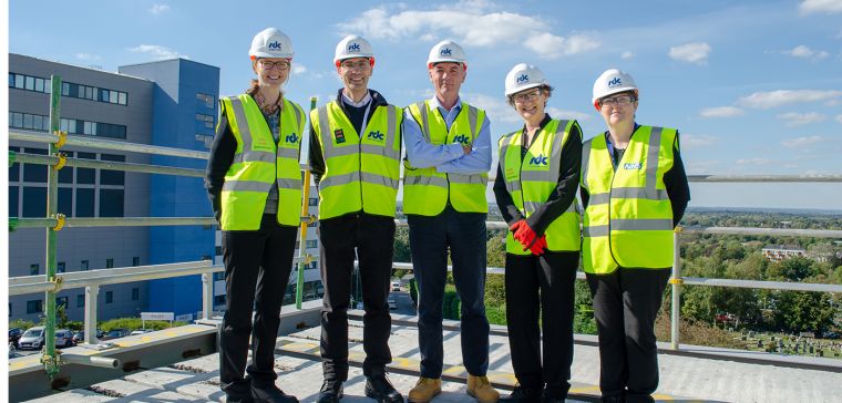
Looking to the future